|
|
|
| |
 |
|
|

|
 推薦指數:
推薦指數:


|
|
- 內容介紹
|
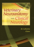
Veterinary Neuroanatomy and Clinical Neurology, 3rd Edition
By Alexander de Lahunta, DVM, PhD and Eric N. Glass, MS, DVM, DACVIM (Neurology)
552 pages
Trim Size 8 1/2 X 10 7/8 in
Copyright 2009
Description
Organized by functional neurologic system, the 3rd edition of this authoritative reference provides the most up-to-date information on neuroanatomy, neurophysiology, neuropathology, and clinical neurology as it applies to small animals, horses, and food animals. Accurate diagnosis is emphasized throughout with practical guidelines for performing neurologic examinations, interpreting examination results, and formulating effective treatment plans. In-depth disease descriptions, color images, and video clips reinforce important concepts and assist with diagnosis and treatment.
Reviews
"The third edition really does cater for everyone; undergraduates, practitioners and specialist neurologists will all find this book an absolute delight. (...) The result is an outstanding combination of neuroanatomy, neuropathology and clinical neurology. A major innovation is the inclusion of text to compliment a series of video clips available for view on a Cornell website. The system works really well, drawing on the authors' vast archive of examples of clinical conditions in all domestic species. There are 382 video clips available, each one with relevant text in the book. (...) The attention to detail in this book is truly impressive and, if the reader wants further information on any topic, there is a comprehensive list of references at the end of each chapter. Much more could be said about this great book but I recommend buying it for oneself; it will be money well-spent."
- The European Journal of Companion Animal Practice, April 2009
Key Features
Expert authors bring more than 50 years of experience in veterinary neuroanatomy and clinical neurology to this book — Dr. Alexander DeLahunta and Dr. Eric Glass offer their unique insights from both academic and practitioner perspectives.
Disease content is presented in a logical case study format with three distinct parts:
Description of the disorder
Neuroanatomic diagnosis (including how it was determined, the differential diagnosis, and any available ancillary data)
Course of the disease (providing final clinical or necropsy diagnosis and a brief discussion of the syndrome)
New to this Edition
More than 600 full-color photographs and line drawings, plus approximately 150 high-quality radiographs, visually reinforce key concepts and assist in reaching accurate diagnoses.
The book comes with free access to 370 video clips on Cornell University’s website that directly correlate to the case studies throughout the book and clearly demonstrate nearly every recognized neurologic disorder.
High-quality MR images of the brain are presented alongside correlating stained transverse sections for in-depth study and comparison.
Vivid photos of gross and microscopic lesions clearly illustrate the pathology of many of the disorders presented in the book
Table of Contents
1. Introduction
2. Neuroanatomy by Dissection
3. Development of the Nervous System: Malformation
4. Cerebrospinal Fluid and Hydrocephalus
5. Lower Motor Neuron: Spinal
6. Lower Motor Neuron: General Somatic
7. Lower Motor Neuron: General Visceral Efferent System
8. Upper Motor Neuron
9. General Sensory Systems:
General Prioprioception-GP
General Somatic Afferent-GSA
10. Small Animal Spinal Cord Disease
11. Large Animal Spinal Cord Disease
12. Vestibular System: Special Proprioception (SP)
13. Cerebellum
14. Visual System
15. Auditory System: Special Somatic Afferent System
16. Visceral Afferent Systems
17. Non-Olfactory Rhinencephalon: Limbic System
18. Seizure Disorders: Narcolepsy
19. Diencephalon
20. The Neurologic Examination
21. Case Descriptions
|
|
|

