|
|
|
| |
 |
|
|

|
 推薦指數:
推薦指數:


|
|
- 內容介紹
|
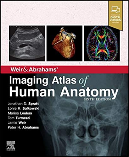
Weir & Abrahams' Imaging Atlas of Human Anatomy 6th Edition
by Jonathan D. Spratt MA (Cantab) FRCS (Eng) FRCR (Author), Lonie R Salkowski MD (Author), Marios Loukas MD PhD (Author), Tom Turmezei BMBCh MA MPhil FRCR (Author)
Publisher : Elsevier; 6th edition
Language : English
Paperback : 288 pages
ISBN-10 : 070207926X
ISBN-13 : 978-0702079269
Item Weight : 2.31 pounds
2021
Imaging is ever more integral to anatomy education and throughout modern medicine. Building on the success of previous editions, this fully revised sixth edition provides a superb foundation for understanding applied human anatomy, offering a complete view of the structures and relationships within the whole body, using the very latest imaging techniques.
All relevant imaging modalities are included, from plain radiographs to more advanced imaging of ultrasound, CT, MRI, functional imaging and angiography. Coverage is further enhanced by a carefully selected range of BONUS electronic content, including clinical photos and cases, ultrasound videos, labelled radiograph ‘slidelines’, cross-sectional imaging stacks and test-yourself materials. Uniquely, key syllabus image sets are now highlighted throughout to aid efficient study, as well as the most common, clinically important anatomical variants that you should be aware of.
This superb package is ideally suited to the needs of medical students, as well as radiologists, radiographers and surgeons in training. It will also prove invaluable to the range of other students and professionals who require a clear, accurate, view of anatomy in current practice.
Fully revised legends and labels and new high-quality images–featuring the latest imaging techniques and modalities as seen in clinical practice
Covers the full variety of relevant modern imaging–including cross-sectional views in CT and MRI, angiography, ultrasound, fetal anatomy, plain film anatomy, nuclear medicine imaging and more – with better resolution to ensure the clearest anatomical views
Core syllabus image sets now highlighted throughout–to help you focus on the most essential areas to excel on your course and in examinations
Unique summaries of the most common, clinically important anatomical variants for each body region–reflects the fact that around 20% of human bodies have at least one clinically significant variant
New orientation drawings–to help you understand the different views and the 3D anatomy of 2D images, as well as the conventions between cross-sectional modalities
Ideal as a stand-alone resource or in conjunction with Abrahams’ and McMinn’s Clinical Atlas of Human Anatomy–where new links help put imaging in the context of the dissection room
Now a more complete learning package than ever before, with superb BONUS electronic enhancements embedded within the accompanying eBook, including:
Labelled image ‘stacks’–that allow you to review cross-sectional imaging as if using an imaging workstation
Labelled image ‘slidelines’–showing features in a full range of body radiographs to enhance understanding of anatomy in this essential modality
Self-test image ‘slideshows’ with multi-tier labelling–to aid learning and cater for beginner to more advanced experience levels
Labelled ultrasound videos–bring images to life, reflecting this increasingly clinically practiced technique
Questions and answers accompany each chapter–to test your understanding and aid exam preparation
34 pathology tutorials–based around nine key concepts and illustrated with hundreds of additional pathology images, to further develop your memory of anatomical structures and lead you through the essential relationships between normal and abnormal anatomy
High-yield USMLE topics–clinical photos and cases for key topics, linked and highlighted in chapters
|
|
|

