|
|
|
| |
 |
|
|

|
-
推薦指數:


|
|
- 內容介紹
|
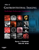
Atlas of Gastrointestinal Imaging: Radiologic-Endoscopic Correlation
By Perry J. Pickhardt, MD and Glen M. Arluk, MD
Approx. 512 pages Approx. 3400 illustrations (950 in full color)
Trim size 8 1/2 X 10 7/8 in
Copyright 2007
Description
This text provides unique and invaluable, cutting-edge information on the latest gastrointestinal imaging techniques. Dr. Pickhardt – a leading researcher in the field of “virtual colonoscopy” – guides you through the use of this new technique as well as all other imaging approaches for the entire GI tract. What’s more, unique correlations between radiology and endoscopy studies help you to see how the findings compare for every diagnosis. An organization by differential diagnosis speeds you to answers to your clinical questions, and hundreds of spectacular color digital-quality images present all of the visual assistance you need to make an accurate diagnosis for both common and uncommon gastrointestinal disorders.
Key Features
View the correlations between radiologic and endoscopic imaging findings with the aid of hundreds of side-by-side images.
Access key, clinically relevant information quickly through an easy-to-read, bulleted format.
Apply the practice-proven methods of Dr. Perry J. Pickhardt, a top researcher in gastrointestinal radiology.
Gain an accurate picture of all diseases and disorders – as well as key anatomic areas – thanks to a full-color format and color anatomic images.
Table of Contents
1. THE ESOPHAGUS
1.1 ESOPHAGEAL TUMORS
Adenocarcinoma
Squamous Cell Carcinoma
Mesenchymal Tumors
Other Esophageal Neoplasms
Non-neoplastic Lesions
1.2 GASTROESOPHAGEAL REFLUX DISEASE (GERD)
Reflux Esophagitis
Peptic Strictures
Barrett’s Esophagus
1.3 ESOPHAGITIS (NON-GERD)
Infectious Esophagitis
Drug-Induced, Caustic, and Radiation Injury
Other Esophagitides
1.4 ESOPHAGEAL DYSMOTILITY DISORDERS
Achalasia
Diffuse Esophageal Spasm
Scleroderma (Progressive Systemic Sclerosis)
1.5 OTHER ESOPHAGEAL CONDITIONS
Vascular Lesions
Pharyngoesophageal Diverticula
Duplication Cysts
Mechanical Injury
Foreign Body Impaction
Esophageal Fistulas
Intramural Pseudodiverticulosis
Rings, Webs, and Stenosis
Vascular Rings and Slings
Hypopharyngeal Disease
2. THE STOMACH
2.1 GASTRIC TUMORS
Mucosal Polyps
Adenocarcinoma
Lymphoma
Mesenchymal Tumors
Other Submucosal Tumors
2.2 GASTRITIS AND GASTROPATHY
Peptic Ulcer Disease
Reactive Gastropathies
Zollinger-Ellison Syndrome
Infectious Gastritis
Granulomatous Diseases
Ménétrier’s Disease
Other Inflammatory Conditions
2.3 OTHER GASTRIC CONDITIONS
Vascular Lesions
Gastric Hernias
Gastric Fistulas
Heterotopic Pancreatic Rest
Duplication Cysts
Gastric Bezoars
Gastric Volvulus
Gastric Diverticula
Complications of Percutaneous Endoscopic Gastrostomy
3. THE DUODENUM
3.1. DUODENAL TUMORS
Mucosal Neoplasms
Submucosal Tumors
Non-neoplastic Lesions
3.2 INFLAMMATORY CONDITIONS
Peptic Ulcer Disease
Duodenitis
3.3 OTHER DUODENAL CONDITIONS
Vascular Lesions
Aortoduodenal Fistula
Duodenal Diverticula
Duodenal Duplication Cyst
Infiltrative Diseases
4. THE MESENTERIC SMALL BOWEL
4.1 SMALL BOWEL TUMORS
Adenocarcinoma
Carcinoid Tumor
Lymphoma
Mesenchymal Tumors
Metastatic Disease
4.2 ENTERITIS
Crohn’s Disease
Infectious Enteritis
Other Inflammatory Conditions
4.3 OTHER SMALL BOWEL CONDITIONS
Small Bowel Obstruction
Mesenteric Ischemia
Small Bowel Herniation
Celiac Disease
Small Bowel Diverticula
Malrotation
Small Bowel Wall Thickening
Small Bowel Perforation
Vascular Ectasia
5. THE COLON AND RECTUM
5.1 COLORECTAL POLYPS AND MASSES
Benign Mucosal Neoplasms
Non-neoplastic Mucosal Lesions
Submucosal Lesions
Colonic Adenocarcinoma
Rectal Adenocarcinoma
Other Colorectal Tumors
CTC Diagnostic Tools
CTC Pitfalls
5.2 COLITIS
Ulcerative Colitis
Crohn’s Disease
Infection
Ischemia
Other Colitides
5.3 COLONIC DIVERTICULAR DISEASE
Diverticulosis
Acute Diverticulitis
Diverticular Fistulas and Strictures
Diverticular Hemorrhage
Giant Sigmoid Diverticulum
5.4 THE APPENDIX
Appendicitis
Appendiceal Tumors
5.5 OTHER COLORECTAL CONDITIONS
Anorectal Disease
Intussusception
Vascular Lesions
Colonic Volvulus
Endometriosis
Pneumatosis Coli
Colonic Hernias
Complications of Colonoscopy
Epiploic Appendagitis
Melanosis Coli
6. THE BILIARY SYSTEM
6.1 BILIARY TUMORS
Cholangiocarcinoma
Gallbladder Carcinoma
Periampullary Tumors
Metastatic Disease
Other Biliary Tumors
6.2 BILIARY CALCULI
Cholelithiasis
Choledocholithiasis
Mirizzi’s Syndrome
Biliary-Enteric Fistulas
6.3 INFLAMMATORY DISEASES
Cholecystitis
Primary Sclerosing Cholangitis (PSC Ascending Cholangitis)
Recurrent Pyogenic Cholangitis
AIDS Cholangiopathy
6.4 OTHER BILIARY CONDITIONS
Choledochal Cysts
Caroli’s Disease
Adenomyomatosis
Porcelain Gallbladder
Biliary Leak
Other Causes of Stricture and Obstruction
Hemobilia
7. THE PANCREAS
7.1 PANCREATIC NEOPLASMS
Ductal Adenocarcinoma
Mucinous Cystic Neoplasms
Intraductal Papillary Mucinous Neoplasm
Serous Cystadenoma
Islet Cell Tumors
Solid-Pseudopapillary Tumor
Metastatic Disease
Other Pancreatic Neoplasms
7.2 PANCREATITIS
Acute Pancreatitis
Chronic Pancreatitis
Pancreatic Pseudocysts
Autoimmune Pancreatitis
7.3 OTHER PANCREATIC CONDITIONS
Pancreas Divisum
Annular Pancreas
Simple Pancreatic Cysts
Pancreatic Trauma
8. THE LIVER
8.1 NEOPLASTIC LESIONS
Hepatocellular Carcinoma
Metastatic Disease
Cavernous Hemangioma
Hepatocellular Adenoma
Intrahepatic (Peripheral) Cholangiocarcinoma
Other Hepatic Tumors
8.2 NON-NEOPLASTIC LESIONS
Focal Nodular Hyperplasia
Benign Hepatic Cysts
Hepatic Abscess
Vascular Findings
8.3 OTHER HEPATIC CONDITIONS
Cirrhosis and Portal Hypertension
Budd-Chiari Syndrome
Fatty Liver Disease (Hepatic Steatosis)
Hemochromatosis
The Hyperdense Liver on CT
Post-transplant Complications
|
|
|

