|
|
|
| |
 |
|
|

|
 推薦指數:
推薦指數:


|
|
- 內容介紹
|
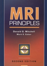
MRI Principles 2e, 2nd Edition
Donald G. Mitchell MD, Professor of Radiology, Director, Division of Magnetic Resonance Imaging, Thomas Jefferson University Hospital, Philadelphia, PA
Mark Cohen PhD, Brain Mapping Division, UCLA School of Medicine, Los Angeles, CA
ISBN 0721600247 · Hardback
Saunders · Published February 2004
The New Edition of this popular, practical resource offers a lucid introduction to the principles of MRI, explaining in plain language the general principles of magnetism and nuclear magnetic resonance induction, and how this phenomenon can be used to generate and manipulate images for clinical use. A wealth of high-quality illustrations, complemented by concise text, enable readers to gain a solid understanding without requiring in-depth knowledge of physics and mathematics. And, each lists of essential points at the end of each chapter enable readers to test and hone their knowledge
Features
Provides a clear understanding of the principles of MRI without assuming specialized knowledge of mathematics and physics.
Features a practical, clinical focus that reflects current practice standards.
Discusses how protocols change with each patient and procedure.
Offers comparative examples, showing an image at different MRI parameters.
What's New
Incorporates new techniques for functional and physiologic imaging
Presents new techniques such as parallel functional imaging and a new method for filling k-space
Offers fresh perspectives from physicist and expert in neuroimaging, Mark Cohen, PhD
Contents
MITCHELL AND COHEN: MRI PRINCIPLES, 2/E
TABLE OF CONTENTS:
CHAPTER 1: What is Magnetic Resonance Imaging?
CHAPTER 2: From Protons to Images
CHAPTER 3: Proton Environments and TI Relaxation
CHAPTER 4: Transverse Magnetization and T2 Contrast
CHAPTER 5: Chemical Shift
CHAPTER 6: Spatial Localization: Magnetic Field Gradients
CHAPTER 7: K-Space: A Graphic Guide
CHAPTER 8: Image Acquisition: Pulse Sequences
CHAPTER 9: Signal-to-Noise Ratio and Spatial Resolution
CHAPTER 10: Acquisition Time Reconsidered
CHAPTER 11: Receiver Coils
CHAPTER 12: Magnetic Field Strength
CHAPTER 13: Motion-Induced Artifacts
CHAPTER 14: Pulse Sequences: Gradient Echo and Spin Echo
CHAPTER 15: Preparatory Pulses, Including Fat Suppression
CHAPTER 16: Multiecho Techniques
CHAPTER 17: Strategies of Fast Imaging
CHAPTER 18: Ti-Weighted Pulse Sequences
CHAPTER 19: T2-Weighted Pulse Sequences
CHAPTER 20: Intermediate-Weighted Pulse Sequences
CHAPTER 21: Intravenous Water-Soluble Contrast Agents
CHAPTER 22: Particulate and Oral Contrast Agents
CHAPTER 23: Contrast-Enhanced MR Angiography
CHAPTER 24: Imaging of Blood Flow
CHAPTER 25: Perfusion and Diffusion Techniques
CHAPTER 26: Artifacts
CHAPTER 27: Cardiac MRI Techniques
CHAPTER 28: Clinical MRI Techniques
|
|
|

