|
|
|
| |
 |
|
|

|
 推薦指數:
推薦指數:


|
|
- 內容介紹
|
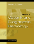
Textbook of Veterinary Diagnostic Radiology 4e
Donald E. Thrall, DVM, PhD, Professor of Radiology, Department of Anatomy, Physiological Science and Radiology, North Carolina State University, College of Veterinary Medicine, Raleigh, NC
ISBN 0721688209 · Hardback · 480 Pages · 885 Illustrations
W.B. Saunders · Forthcoming Title (March 2002)
This text contains information on diagnostic radiology of canine, feline and equine species. Also included are chapters on ultrasound, CT and MRI. Designed to present diagnostic radiology in a user-friendly approach, it starts with the physics of radiology and then moves into interpretation of the radiographs. Radiographic anatomy helps the reader formulate a diagnosis. Questions at the end of each chapter, with answers at the end of the book, reinforce important concepts and ensure that readers understand one concept before progressing to the next.
Features
In-depth coverage of the radiographic anatomy of the dog, cat, and horse enables the reader to recognize the more frequently radiographed regions.
A body systems approach presents information in a logical progression and makes topics easy to find.
Discussion of the physics of radiology, CT, and MRI gives readers a better understanding of the radiographic process.
Information on radiation safety highlights safety measures associated with ionizing radiation.
Questions with answers reinforce important points and facilitate a better understanding of diagnostic radiology.
High-quality radiographs provide excellent examples of diagnostic criteria
What's New
* Coverage of ultrasound physics is incorporated into this edition.
Interpretation paradigms - modules set up to demonstrate how to properly interpret a radiograph - are included for:
* the axial skeleton
the appendicular skeleton - canine & feline
the appendicular skeleton - equine
the tarsus
the thorax - canine and feline
the abdomen - canine and feline
Contents
Section 1. Physics and Principles of Interpretation
Physics of Diagnostic Radiology, Radiology Protection, and Darkroom Theory
Ultrasound Physics
Physics of Computed Tomography and Magnetic Resonance Imagine
Visual Perception
Introduction to Radiographic Interpretation
Section 2. Axial Skeleton
Interpretation Paradigms for the Axial Skeleton
The Cranial and Nasal Cavities - Canine and Feline
Equine Nasal Passages and Sinuses
The Vertebrae - Canine and Feline
Intervertebral Disc Disease - Canine and Feline
The Equine Vertebral Column
Section 3. Appendicular Skeleton - Canine and Feline
Interpretation Paradigms for the Appendicular Skeleton - Canine and Feline
Diseases of the Immature Skeleton - Small Animal
Fracture Healing and Complications
Bone Tumors versus Bone Infection
Radiographic Signs of Joint Disease
Section 4. Appendicular Skeleton - Equine
Interpretation Paradigms for the Appendicular Skeleton - Equine
The Stifle
The Tarsus
The Carpus
The Metacarpus and Metatarsus
The Metacarpophalangeal (Metatarsophalangeal) Articulation
The Phalanges
The Navicular Bone
Section 5. Neck and Thorax - Companion Animals
Interpretation Paradigms for the Canine and Feline Thorax
The Larynx, Pharynx and Trachea
The Esophagus
The Thoracic Wall
The Diaphragm
The Mediastinum
The Pleural Space
The Heart and Great Vessels
The Pulmonary Vasculature
The Lung
Section 6. Neck and Thorax - Equidae
The Larynx, Pharynx and Trachea
The Equine Lung
The Pleural Space
Section 7. Abdomen - Companion Animals
Interpretation Paradigms for the Abdomen - Canine and Feline
Abdominal Masses
The Peritoneal and Retroperitoneal Spaces
The Liver and Spleen
The Kidneys and Ureters
The Urinary Bladder
The Urethra
The Prostate Gland
The Uterus, Ovaries and Testes
The Stomach
The Small Bowel
The Large Bowel
Section 8. Radiographic Anatomy
50. Radiographic Anatomy of the Dog and Horse
|
|
|

