|
|
|
| |
 |
|
|

|
 推薦指數:
推薦指數:


|
|
- 內容介紹
|
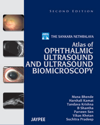
The Sankara Nethralaya Atlas of Ophthalmic Ultrasound and Ultrasound Biomicroscopy
by: Muna Bhende, Harshali Kamat, Tandava Krishna, B Shantha, Parveen Sen, Vikas Khetan, Suchitra Pradeep
ISBN: 9789350905357
Edition: 2/e, 2013
Pages: 440
Size: 8.5" X 11"
Price: Rs. 2495
Cover Type: Hard Back
The first edition of The Sankara Nethralaya Atlas of Ophthalmic Ultrasound was intended to be both a teaching and practical reference tool for the ophthalmologist. Though more sophisticated imaging techniques in certain areas of ophthalmology have evolved over the past years, ultrasonography is still a useful tool that can be done in a clinic by the ophthalmologist. While updating some of the chapters of the first edition with more images and examples of representative cases, the authors have retained the original outline and pattern.
In this second edition The Sankara Nethralaya Atlas of Ophthalmic Ultrasound and Ultrasound Biomicroscopy, authors have now included an entirely new section on ultrasound biomicroscopy that has widened the scope of this atlas to include anterior segment imaging as well. With text being kept to a minimum, the salient clinical and ultrasound features have been described with practical tips for technique, interpretation, and implications on management. Where possible, authors have provided corresponding color photographs, pathology images as well as other diagnostic reports, such as CT and MRI. The combined experience of ophthalmologists from almost all the ophthalmic subspecialties has resulted in a large collection of cases both common and rare, where ultrasound imaging has been a valuable diagnostic aid.
Key Features The second edition of the Sankara Nethralaya atlas of ophthalmic ultrasound, while retaining its original layout and flavor, has considerably widened its scope to include anterior segment imaging using ultrasound biomicroscopy. The original chapters which involved imaging of the posterior segment and orbit have been updated with addition of many new images, though the techniques of evaluation have remained unchanged over the years. The new section on ultrasound biomicroscopy includes common and rare conditions which involve the anterior segment with corresponding photographs where possible. Practical issues as to when to perform the test in a given condition, what to look for and the implications on treatment have been dealt with in a brief fashion. This atlas aims to educate the novice and at the same time refresh the knowledge of the qualified ophthalmologist in one of the segments in the always fascinating field of ophthalmic imaging - ultrasonography.
|
|
|

