|
|
|
| |
 |
|
|

|
 推薦指數:
推薦指數:


|
|
- 內容介紹
|
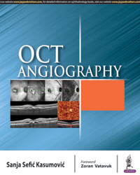
OCT Angiography
Author:Sanja Sefic Kasumovic MD PhD
Item Weight : 1.95 pounds
Hardcover : 200 pages
ISBN-10 : 9352703316
ISBN-13 : 978-9352703319
Dimensions : 8.5 x 11 inches
Publisher : Jaypee Brothers Medical Pub; 1/e edition
Language: : English
2019
Quick Overview
The book OCT Angiography is a handwriting which presents a good example of establishing a correlation with world literature in the field of new, sophisticated ophthalmology diagnostics. It represents a concise and sufficiently detailed monography on the new OCTA diagnostic method in ophthalmology. Every single image includes descriptions and comments, which gives the material a great value and worthiness. Some of pathological cases are presented with ten to fifteen individual images and figures, aiming to help the reader properly read and analyse the patient from beginning to the end. Also, images are organized so the reader can easily compare and understand correlation between them. Documented findings give the physician a possibility of careful comparison with earlier findings of the classic OCT. Author has very skillfully introduced a reader into anatomy, physiology and pathology of the most common diseases of the posterior ocular segment. The book gives also a very good overview of optic nerve pathology and OCTA findings. Monography OCT Angiography completes the requirements of educational literature from the domain of diagnostics the posterior eye segment and can serve ophthalmologists, residents of ophthalmology as well as medical students.
Key Features
• The book OCT angiography represents a handy guide through the OCTA technique while illustrating and explaining various retinal diseases as well as pathology of the optic nerve
• The reader is provided with a short description of epidemiology, etiology, clinical signs and symptoms, as well as diagnostic and treatment possibilities for every entity described
• The book is full of illustrations, colored figures and maps of every single case from everyday clinical practice and it provides a detailed step-by-step explanation below the every figure, with the aim of better insight and logical understanding of the retinal pathology
• It is easy-to-read material, intended for medical students, residents of ophthalmology and ophthalmology specialists who are willing to expand their knowledge and to flow into the new technique of retina vascularization imaging
• Every single case is represented by multiple images, which are arranged one-by-one, aiding in easy pattern recognition, diagnosis and monitoring of patients
• It is updated diagnostic guidance for glaucoma experts who are willing to find out the real semiology of glaucoma development.
|
|
|

