|
|
|
| |
 |
|
|

|
 推薦指數:
推薦指數:


|
|
- 簡介
|
ISBN 9789351525325
Edition 1/e
Publish Year 2015
Pages 108
Size 8.5"x11"
Cover Type Hard Back
With CD/DVD No
|
|
- 內容介紹
|
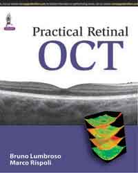
OCT highlights retinal and choroidal alterations in morphology, structure and reflectivity and facilitates the study of the various retinal layers, both separately and globally. The images presented in this manual were recorded mainly with Optovue OCT (RTVue and XR Avanti OCT), and also with Heidelberg Spectralis. These instruments are both reliable and easy to use. There are high resolution optical sections, in sagittal view with the choroid visible to the sclera, with or without image averaging, or in frontal view, "en face", adapted the curvature of the fundus and other 3D images. Illustrated with drawings, outlines, and sagittal and frontal (“en face”) OCT images, this concise manual intends to show how to read and interpret OCTs, documenting and diagnosing the most common retinal pathologies. Simplified outlines are provided to facilitate the classification of morphological alterations.
|
|
|

