|
|
|
- 您 可 以 信 任 的 醫 學 圖 書 智 庫 |
|
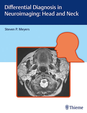
Differential Diagnosis in Neuroimaging: Head and Neck 2017
 Differential Diagnosis in Neuroimaging: Head and Neck 2017
-放射線科
- 編號: 0----001626234752
- 作者: Steven Meyers
- 原價-7215 - (熱賣價)5920 - 節省 ↓18%
- 加入購物車 提交
推薦指數:
- 內容介紹
Differential Diagnosis in Neuroimaging: Head and Neck
by Steven Meyers (Editor)
Hardcover: 664 pages
Publisher: Thieme; 1 edition
Language: English
ISBN-10: 1626234752
ISBN-13: 978-1626234758
Product Dimensions: 8.5 x 1.5 x 11 inches
2017
Authored by renowned neuroradiologist Steven P. Meyers, Differential Diagnosis in Neuroimaging: Head and Neck is a stellar guide for identifying and diagnosing head and neck disease based on location and neuroimaging results. The succinct text reflects more than 25 years of hands-on experience gleaned from advanced training and educating residents and fellows in radiology, neurosurgery, and otolaryngology. The high-quality MRI and CT scans have been collected over Dr. Meyers's lengthy career, presenting an unsurpassed visual learning tool.
The distinctive 'three-column table plus images' format is easy to incorporate into clinical practice, setting this book apart from larger, disease-oriented radiologic tomes. This layout enables readers to quickly recognize and compare abnormalities based on more than 1,500 high-resolution images. Chapters cover skull imaging, temporal bone imaging, orbital imaging, paranasal imaging, suprahyoid neck imaging, and infrahyoid neck imaging, for a full spectrum of head and neck pathologies.
Key Highlights
•Tabular columns organized by anatomical abnormality include neck, facial, and skull based imaging findings and a summary of key clinical data that correlate to the images
•Congenital/developmental and acquired abnormalities including solitary or multiple orbital lesions; and solitary, multifocal, or diffuse sinonasal disease
•Abnormalities of the skull, craniovertebral junction, tempormandibular joint, infrahyoid neck, anterior and posterior cervical space, perivertebral space, and brachial plexus
This visually rich resource is a must-have diagnostic tool for residents, fellows, and practitioners in radiology, otolaryngology-head and neck surgery, and neurosurgery. The highly practical format makes it ideal for daily rounds, as well as a robust study guide for physicians preparing for board exams.
|
|
|
|
|
└ 返回上一頁
|
|

